100% satisfaction guarantee Immediately available after payment Both online and in PDF No strings attached
logo-home
Summary IB Biology Topic 6: Human Physiology £4.44   Add to cart

Summary

Summary IB Biology Topic 6: Human Physiology

 30 views  0 purchase
  • Module
  • Institution
  • Book

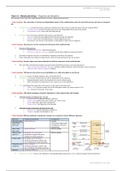
Detailed objective-by-objective summary notes for Topic 6: Human Physiology for IB Biology HL. Contains information on everything you need to know from 6.1 to 6.5, according to each understanding, application or skill. Written by a IB HL Biology student who graduated with a 45/45.

Preview 3 out of 16  pages

  View example

Getting your document ready...

mobile-preview
  • Unknown
  • March 5, 2019
  • 16
  • 2016/2017
  • Summary
avatar-seller
IB TOPIC 6 | HUMAN PHYSIOLOGY
2016 | SYJ0014


Topic 6.1: Human physiology – Digestion and absorption
The structure of the wall of the small intestine allows it to move, digest and absorb food.

• Understanding: The contraction of circular and longitudinal muscle of the small intestine mixes the food with enzymes and moves it along the
gut.

 Smooth muscles: muscles that exerts continuous moderate force that consists of relatively short cells not elongated fibres
 Circular muscle: constricts the gut to prevent it from being pushed back towards the mouth
 Longitudinal muscle: moves the food along the gut

 Peristalsis: wave of muscle contraction that occurs in one direction
 Function: churns the semi-digested food with the enzyme for digestion
 Purpose: moves the food along the gut to initiate digestion process
 Food moves very slowly in the intestine via peristalsis to allow time for digestion

• Understanding: The pancreas secretes enzymes into the lumen of the small intestine.

 Function of the pancreas:
 Secretion of hormone: insulin and glucagon
 Secretion of digestive enzymes: amylase (starch), lipase (lipids), proteases (proteins)

 Secretion of digestive enzymes is controlled by hormones secreted by the stomach
 Ducts within pancreas merge into larger ducts to form one large pancreatic duct for secretion

• Understanding: Enzymes digest most macromolecules in food into monomers in the small intestine.

 The wall of the small intestine produce enzymes (both immobilized enzymes and as intestinal juice)
 Types of enzymes produced by intestine: nuclease, maltase (maltose), lactase (lactose), sucrase (sucrose)
 Substance that cannot be digested: cellulose

• Understanding: Villi increase the surface area of epithelium over which absorption is carried out.

 Absorption: process of taking substance into cells and the blood
 Assimilation: conversion of nutrients into solid or liquid parts of the organism
 Digested materials need to be absorbed to be used in cell processes
 Rate of absorption is proportional to the surface area of the epithelium

 Villi: small finger-like projection of the mucosa on the inside in intestinal wall
 Surface area: increases surface area of the absorption surface (by about a factor of 10)
 Size: between 0.5mm and 1.5mm long

• Understanding: Villi absorb monomers formed by digestion as well as mineral ions and vitamins.

 Absorbed products of digestion by t he villi:
 Monosaccharides: glucose, fructose, galactose
 Amino acids: broken down from twenty; any of the twenty
 Lipids: fatty acids, glycerides and glycerol
 Nucleic bases: from digestion of nucleotides

 Absorbed products required by the body by the villi:
 Mineral ions: calcium, potassium, sodium etc.
 Vitamins: ascorbic acid (vitamin C) etc.

• Understanding: Different methods of membrane transport are required to absorb different nutrients.

Molecule Transport mechanism Transported
Monoglyceride Simple diffusion Lacteal
Long chain (units forms triglyceride
fatty acid in epithelial cells and is
transported)
Short chain Simple diffusion Blood capillary
fatty acids
Glucose and Secondary active transport Blood capillary
galactose (facilitated coupled with active
building of sodium gradient)
Amino acids Secondary active transport
(facilitated transport coupled with
active building of sodium gradient)
Dipeptides and Active transport
tripeptides (facilitated transport coupled with
active building of hydrogen gradient)
Fructose Facilitated diffusion

 Lipoprotein complex: forms in the epithelium when phospholipids and proteins covers reformed triglyceride and cholesterol


LAST EDITED 2017-03-16 | 1

, IB TOPIC 6 | HUMAN PHYSIOLOGY
2016 | SYJ0014


• Application: Processes occurring in the small intestine that result in the digestion of starch and transport of the products of digestion to the
liver.

 Starch: a long chain of α-glucose molecules that consists of 1,4 bonds and 1,6 bonds (specific in amylopectin)

Enzyme Target bond Target reactant Product
Amylase 1,4 α-glucose bonds Starch (chain of at least four α-glucose molecules) Maltase, maltotriase, dextrinase
Maltase 1,4 α-glucose bonds Maltose (fragment two α-glucose) α-glucose
Maltotriase 1,4 α-glucose bonds Maltotriose (fragment of three α-glucose) α-glucose
Dextrinase 1,6 α-glucose bonds Dextrin (fragment of amylopectin containing 1,6 bonds) α-glucose

 Process of glucose transport and storage in liver:
 Villus epithelium: glucose is absorbed into villus epithelium by co-transport with sodium ions
 Blood capillary: glucose enters the porous blood capillary through diffusion
 Venules: glucose in blood capillary flows through to the venules in the sub-mucosa
 Hepatic portal vein: venules then flows to the hepatic portal vein to the liver
 Liver: excess glucose is absorbed and stored by glycogen or vice versa

• Application: Use of dialysis tubing to model absorption of digested food in the intestine.

 Dialysis tube: semi-permeable tube that allows molecules of smaller size than the pore to pass through

 Method of observation in function of intestine through dialysis tubes:
 Dialysis tube filling: dialysis tube is filled with solution of starch and glucose
 Dialysis tube immersion: dialysis tube is then immersed into a beaker of distilled water
 Observation and record: after one hour, only glucose is present in water

• Skill: Production of an annotated diagram of the digestive system.

Structure Function
Mouth Control of eating and swallowing; mechanical digestion of food and initiating chemical
digestion by mixing food with saliva
Esophagus Movement of food from mouth to stomach through peristalsis
Stomach Killing of foreign bacteria by mixing with water and acid and initial stage of protein digestion
Pancreas Secretion of enzymes (amylase, protease, lipases)
Liver Secretion of surfactants bile to digest lipids and for neutralization
Gall bladder Storage and regulation of bile release
Small intestine Production of enzymes, final stages of digestion (of carbohydrates, lipids, proteins and
nucleic acid) and stages of absorption of nutrients
Large intestine Re-absorption of water, further digestion by symbiotic bacteria

• Skill: Identification of tissue layers in transverse sections of the small intestine viewed with a microscope or in a micrograph.

Structure Function
Epithelial layer Selective absorption of nutrients
Mucosa Lining of the small intestine that produces mucus that protects cells
Submucosa Tissue layer containing blood and lymph vessels
Circular muscle Cause waves of contraction; peristalsis
Longitudinal muscle Moves food along the gut and aids peristalsis

• Nature of science: Use models as representations of the real world—dialysis tubing can be used to
model absorption in the intestine.

• Utilization: Some hydrolytic enzymes have economic importance, for example amylase in production of sugars from starch and in the brewing of beer.
• Guidance: Students should know that amylase, lipase and an endopeptidase are secreted by the pancreas. The name trypsin and the method used to
activate it are not required.
• Guidance: Students should know that starch, glycogen, lipids and nucleic acids are digested into monomers and that cellulose remains undigested.
• Guidance: Tissue layers should include longitudinal and circular muscles, mucosa and epithelium.


LAST EDITED 2017-03-16 | 2

, IB TOPIC 6 | HUMAN PHYSIOLOGY
2016 | SYJ0014


Topic 6.2: Human physiology – The blood system
The blood system continuously transports substances to cells and simultaneously collects waste products.

• Understanding: Arteries convey blood at high pressure from the ventricles to the tissues of the body.

 Arteries: vessels that convey blood at high pressure and speed away from the heart to the body
 Speed: high speed (10-40 cm/s)
 Pressure: high pressure (80-120 mmHg)

 Blood moves in pulses due to expanding and recoiling when pressure rises and falls
 Each organ is supplied by one or more arteries

• Understanding: Arteries have muscle cells and elastic fibres in their walls.

 Arteries have muscle cells and elastic fibres to maintain toughness of the walls
 Elastin fibres: stores energy that stretches them at the peak of each pumping cycle; recoiling
help propel blood down artery
 Muscle cells: contraction determines the pulsating movement

Structure Composition Function
Tunica externa Elastin and collagen Maintains integrity of muscles
Tunica media Smooth muscles Assist increasing and decreasing the pressure of the artery for
pulsating movement
Tunica intima Endothelium cells Prevent adhesion of blood cells to the walls of vessels

• Understanding: The muscle and elastic fibres assist in maintaining blood pressure between pump cycles.

 Mechanism to maintain blood flow in arteries:
 Ventricle contraction: ventricles contract, increasing blood pressure in arteries
 Artery potential energy storage: this pushes the wall of the artery outward, stretching elastic fibres that stores potential energy
 Blood movement forward: pressure falls at the end of the heartbeat, and the stretched elastic fibres squeeze the blood forward

 Such process saves energy and prevents the minimum pressure from becoming too low

 Systolic blood pressure: peak pressure reached in the artery
 Diastolic blood pressure: lowest pressure reached in the artery

 Vasoconstriction: when the circular muscle contract to narrow the lumen for higher pressure and less blood flow
 Vasodilation: when the circular muscle relaxes to expand the lumen for lower pressure and more blood flow

• Understanding: Blood flows through tissues in capillaries. Capillaries have permeable walls that allow exchange of materials between cells in
the tissue and the blood in the capillary.

 Capillaries: blood vessels that have permeable walls that allow exchange of material between cells and the blood
 Speed: low speed (0.1 cm/s)
 Pressure: moderate pressure (15 mmHg)

 Capillaries are the narrowest blood vessel that reach all cells in body
 Capillaries join arteries (and arterioles) to veins (and venules)

 Tissue fluid: fluid that contain oxygen, glucose and other substances in blood plasma (except proteins) that diffuses in/out of the capillaries
through the permeable wall to flows between the cells in the tissues to allow for exchange of materials

• Understanding: Veins collect blood at low pressure from the tissues of the body and return it to the atria of the heart.

 Veins: vessels that convey blood at low pressure and speed away from the heart to the body
 Speed: moderate speed (5-20 cm/s)
 Pressure: low pressure (<10 mmHg)

 Blood is in much lower pressure and therefore do not need a thick tunica media (muscle layer)
 Most organs are served by one or more vein, and the hepatic portal vein is and exception in its function

 Roles of gravity and skeletal muscle pressure:
 Gravity: blood flow in vein can move along gravity to minimize energy use
 Skeletal muscle pressure: contraction muscle shorter and wider so it squeezes on adjacent veins like a pump

• Understanding: Valves in veins and the heart ensure circulation of blood by preventing backflow.

 Valves: flaps of tissues connected to the muscles that opens in one direction and closes in a backflow
 Valves in veins: veins have a low pressure so often there is a danger of backflow of blood
 Valves in heart: the heart is also in danger of backflow during the cyclic change in pressure


LAST EDITED 2017-03-16 | 3

The benefits of buying summaries with Stuvia:

Guaranteed quality through customer reviews

Guaranteed quality through customer reviews

Stuvia customers have reviewed more than 700,000 summaries. This how you know that you are buying the best documents.

Quick and easy check-out

Quick and easy check-out

You can quickly pay through credit card for the summaries. There is no membership needed.

Focus on what matters

Focus on what matters

Your fellow students write the study notes themselves, which is why the documents are always reliable and up-to-date. This ensures you quickly get to the core!

Frequently asked questions

What do I get when I buy this document?

You get a PDF, available immediately after your purchase. The purchased document is accessible anytime, anywhere and indefinitely through your profile.

Satisfaction guarantee: how does it work?

Our satisfaction guarantee ensures that you always find a study document that suits you well. You fill out a form, and our customer service team takes care of the rest.

Who am I buying these notes from?

Stuvia is a marketplace, so you are not buying this document from us, but from seller Stratiotes_Cirrhilabrus.rubriventralis. Stuvia facilitates payment to the seller.

Will I be stuck with a subscription?

No, you only buy these notes for £4.44. You're not tied to anything after your purchase.

Can Stuvia be trusted?

4.6 stars on Google & Trustpilot (+1000 reviews)

13 documents were sold in the last 30 days

Founded in 2010, the go-to place to buy revision notes and other study material for 15 years now

Start selling
£4.44
  • (0)
  Add to cart